Ultrasound-guided interventionism
How are spinal infiltrations performed?
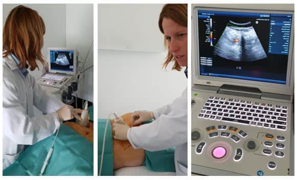
It is essential to adapt to the patient’s needs, with honesty and to offer the patient all the therapeutic alternatives with proven efficacy, surgical and non-surgical, therefore Barcelona Spine Institute has its own Unit of non-invasive therapies, coordinated by Dr. Paola Cavasin, in which innovative techniques such as ultrasound-guided infiltrations are performed, without the need to irradiate the patient.
Ultrasound is an imaging technique based on the emission and reception of ultrasound by a probe. It is mainly used as a diagnostic procedure and puncture guide in different fields of medicine (radiology, cardiology, gynecology…).
The excellent ultrasound visualization of the anatomical structures of the musculoskeletal system has made it possible to adopt new techniques that were not possible before.
Traditionally, interventional techniques are performed by fluoroscopy (X-ray). However, fluoroscopy produces potentially harmful effects for both the patient and the healthcare personnel. In addition, it must be taken into account that fluoroscopy requires specific facilities for its use.
Ultrasound, on the other hand, is non-toxic and, since it does not require specialized facilities, it can be used in the same medical office.
Also, unlike fluoroscopy, ultrasound also allows us to visualize blood vessels, muscles, fasciae and certain cavities or spaces that are very often the origin of the pain we want to treat. In addition, ultrasound expands the diagnostic possibilities by localizing joint effusions, ruptured muscle and tendon fibers, osteoarthritis, cysts and inflammatory processes.
It should be remembered that fluoroscopy consists of the visualization of bone structures and only by using radiological contrast, soft tissues can be observed.
The indication for an interventional technique for the treatment of chronic pain arises when conservative treatment consisting of pharmacological and rehabilitation measures have not been effective.
The identification of the structure we want to treat is achieved with the use of Ultrasound which, in summary, allows the following advantages:
Precise working without the need for irradiation.
Real-time visualization of needle and drug diffusion in our target.
Precisely identify the joints and the superficial and deep musculature to be treated, as well as the neighboring visceral (pleura, kidney) and vascular structures, in order to perform the muscle block with the utmost safety.
In the field of vertebral pathology, the use of ultrasound allows us the guided treatment of:
- Spinal pain of myofascial origin
- Vertebral pain of facet origin
- Pain arising from the sacro-iliac joint
- Sacro-coccygeal and coccygeal joint pain or coccygodynia
Articular and myofascial blocks can be:
- Diagnostics: Local anesthetics such as lidocaine or bupivacaine are used to determine the origin and the affected area of pain.
- Therapeutics: Corticosteroids or botulinum toxin or platelet growth factors may be used for longer lasting relief.
Before performing the technique, a blood test with normal coagulation tests is required.
The blocking cannot be performed if it exists:
- Local or systemic infection.
- Alterations in coagulation.
- Allergy to the drugs used.